Tag Archives: innovation

Graphite for Very High Temperature Reactors (VHTR)

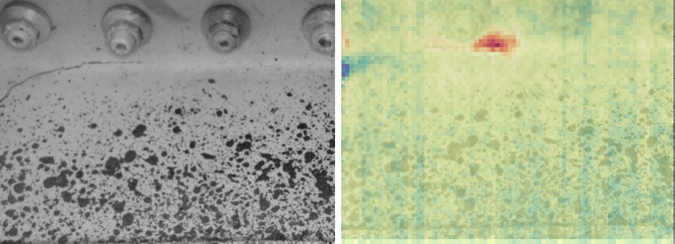
One of the implications of the second law of thermodynamics is that the thermal efficiency of power stations increases with their operating temperature.  Thus, there is a drive to increase the operating temperature in the next generation of nuclear power stations, known as Generation IV reactors.  In one type of Generation IV reactors, known as the Very High Temperature Reactor (VHTR), graphite is designed to be both the moderator for neutrons and a structural element of the reactor.  Although the probability of damage in an accident is extremely low, it is important to consider the consequences of damage causing the core of the reactor to be exposed to air.  In these circumstances, with the core temperature at about 1600°C, the graphite would be exposed to severe oxidation by the air that could change its material properties and ability to function as a moderator and structural element.  Therefore, in recent research, my research group has been working with colleagues at the UK National Nuclear Laboratory (NNL) and at the National Tsing Hua University (NTHU) in Taiwan to conduct experiments on nuclear graphite over a range of temperatures.  Our recently published article shows that all grades of nuclear graphite show increased rates of oxidation for temperatures above 1200°C.  We found that large filler particles using a pitch-based graphite rather than a petroleum-based graphite gave higher oxidation resistance at these elevated temperatures.  This data is likely to be important in the design and operations of the next generation of nuclear power stations.

The work described above was supported by the NTHU-University of Liverpool Dual PhD Programme [see ‘Citizens of the world‘ on November 27th, 2019] and NNL.  This is the fifth, and for the moment last, in a series of posts on recent work published by my research group.  The others are: ‘Salt increases nanoparticle diffusion‘ on April 22nd, 2020; ‘Spatio-temporal damage maps for composite materials‘ on May 6th, 2020; ‘Thinking out of the box leads to digital image correlation through space‘ on June 24th, 2020; and, ‘Potential dynamic buckling in hypersonic vehicle skin‘ on July 1st, 2020.

The image is figure 5: SEM micrographs of the surface of petroleum-based IG-110 graphite samples oxidized at various temperatures from Lo IH, Tzelepi A, Patterson EA, Yeh TK. A study of the relationship between microstructure and oxidation effects in nuclear graphite at very high temperatures.  J. Nuclear Materials. 501:361-70, 2018.

Source:

Lo I-H, Yeh T-K, Patterson EA & Tzelepi A, Comparison of oxidation behaviour of nuclear graphite grades at very high temperatures, J. Nuclear Materials, 532:152054, 2020.

Balancing conscious and unconscious life

Recently, I visited a local artist to choose a painting for a birthday present.  He showed me a pair of small oil paintings in which I had expressed an interest via photographs he had sent me by email.  I agreed to buy both of them and then we drifted into his studio where he showed me the pieces he was working on.  There were many unfinished paintings and he described how difficult it was to finish some of them.  He measured the time taken on some of them in months and, for a few, in years.  I was struck by the similarity with scientists who indulge in slow-motion multi-tasking and switch between research projects in different fields, often leaving something unfinished to focus on something else and then returning to pursue the original research topic [‘Slow-motion multi-tasking leading to productive research‘ on September 19th, 2018].  I suspect both artists and scientists who indulge this approach are looking to achieve ‘a perfect balance of their conscious and unconscious life’ out of which Barbara Hepworth believed ideas are born and realized [see ‘Ideas from a balanced mind‘ on August 24th, 2016].

The studio in the photograph is Barbara Hepworth’s in St Ives, Cornwall.

Thinking out of the box leads to digital image correlation through space

This is the third in a short series of posts on recent engineering research published by my research group.  Actually, two have already been published: ‘Salt increases nanoparticle diffusion‘ on April 22nd, 2020; and ‘Spatio-temporal damage maps for composite materials‘ on May 6th, 2020 and then I got distracted.  This third one arose from the same project as the time-damage maps which was sponsored by the United States Air Force.  The time-damage maps allow us to explore the evolution of failure in complex materials; however, we already know that damage tends to initiate from imperfections or flaws in the microstructure in the material.  New continuous fibre reinforced composite (CFRC) materials based on ceramics are very sensitive to defects or anomalies in their microstructure, such as misalignment of fibres.  However, they are capable of withstanding temperatures in excess of 1500 degrees Centigrade, which offers the opportunity to use them in jet engines or nuclear power plants to help generate energy more efficiently.  Therefore, it is worthwhile investigating effective methods of inspecting their microstructure which we can do either destructively by repetitively polishing away the surface of a sample and viewing it in a microscope, or non-destructively using x-ray tomography.  In both cases, the result is hundreds of ‘images’ containing millions of data values from which it is challenging to extract useful information.  In our work, we have used a little lateral thinking, to show how digital image correlation, usually used to track deformation of structures using multiple images collected over time [see ‘256 shades of grey‘ on January 22nd, 2014] , can be used to track fibres through the multiple images of the layers of the microstructure.  The result is the sort of ‘stick’ diagram in the image showing the orientation of fibres through the sample.  We have demonstrated that our new algorithm was more reliable and 30 times faster than its nearest rival.

The image shows, at the top, a typical stack of images from the microscope of a ceramic matrix composite; and, at the bottom, a plot of 3d profiles of the fibres tracked using the DIC-based method with the fibres orientated nominally at ±45° from the sectioning (x-y) plane shown in red and green colours.

Source:

Amjad K, Christian WJR, Dvurecenska K, Chapman MG, Uchic MD, Przybyla CP & Patterson EA, Computationally efficient method of tracking fibres in composite materials using digital image correlation, Composites Part A, 129:105683, 2020.


Condition-monitoring using infrared imaging

If you have travelled in Asia then you will probably have experienced having your health monitored by infrared cameras as you disembarked from your flight.  It has been common practice in many Asian countries since long before the COVID-19 pandemic and perhaps will become more usual elsewhere as a means of easily identifying people with symptoms of a fever that raises their body temperature.  Since, research has shown that infrared thermometers are slightly more responsive as well as quicker and easier to use than other types of skin surface thermometers [1].  In my research group, we have been using infrared cameras for many years to monitor the condition of engineering structures by evaluating the distribution of load or stress in them [see ‘Counting photons to measure stress‘ on November 18th, 2015 and  ‘Insidious damage‘ on December 2nd, 2015].  In the DIMES project, we have implemented a low-cost sensor system that integrates infrared and visible images with information about applied loads from point sensors, which allows the identification of initiation and tracking of damage in aircraft structures [2].  I reported in December 2019 [see ‘When seeing nothing is a success‘] that we were installing prototype systems in a test-bench at Empa.  Although the restrictions imposed by the pandemic have halted our tests, we were lucky to obtain data from our sensors during the propagation of damage in the section of wing at Empa before lockdown.  This is a landmark in our project and now we are preparing to install our system in test structures at Airbus once the pandemic restrictions are relaxed sufficiently.  Of course, we will also be able to use our system to monitor the health of the personnel involved in the test (see the top image of one of my research team) as well as the health of the structure being tested – the hardware is the same, it’s just the data processing that is different.

The image is a composite showing images from a visible camera (left) and processed data from infrared camera overlaid on the same visible image (right) from inside a wing box during a test at Empa with a crack extending from left to right with its tip surrounded by the red area in the right image.  Each nut in the image is about 20 mm in diameter and a constant amplitude load at 1.25 Hz was being applied causing a wing tip displacement of 80 mm +/- 15 mm.

The University of Liverpool is the coordinator of the DIMES project and the other partners are Empa, Dantec Dynamics GmbH and Strain Solutions Ltd.

The DIMES project has received funding from the Clean Sky 2 Joint Undertaking under the European Union’s Horizon 2020 research and innovation programme under grant agreement No. 820951.


The opinions expressed in this blog post reflect only the author’s view and the Clean Sky 2 Joint Undertaking is not responsible for any use that may be made of the information it contains.

References

[1] Burnham, R.S., McKinley, R.S. and Vincent, D.D., 2006. Three types of skin-surface thermometers: a comparison of reliability, validity, and responsiveness. American journal of physical medicine & rehabilitation, 85(7), pp.553-558.

[2] Middleton, C.A., Gaio, A., Greene, R.J. and Patterson, E.A., 2019. Towards automated tracking of initiation and propagation of cracks in aluminium alloy coupons using thermoelastic stress analysis. Journal of Nondestructive Evaluation, 38(1), p.18.