 If you have travelled in Asia then you will probably have experienced having your health monitored by infrared cameras as you disembarked from your flight. It has been common practice in many Asian countries since long before the COVID-19 pandemic and perhaps will become more usual elsewhere as a means of easily identifying people with symptoms of a fever that raises their body temperature. Since, research has shown that infrared thermometers are slightly more responsive as well as quicker and easier to use than other types of skin surface thermometers [1]. In my research group, we have been using infrared cameras for many years to monitor the condition of engineering structures by evaluating the distribution of load or stress in them [see ‘Counting photons to measure stress‘ on November 18th, 2015 and ‘Insidious damage‘ on December 2nd, 2015]. In the DIMES project, we have implemented a low-cost sensor system that integrates infrared and visible images with information about applied loads from point sensors, which allows the identification of initiation and tracking of damage in aircraft structures [2]. I reported in December 2019 [see ‘When seeing nothing is a success‘] that we were installing prototype systems in a test-bench at Empa. Although the restrictions imposed by the pandemic have halted our tests, we were lucky to obtain data from our sensors during the propagation of damage in the section of wing at Empa before lockdown. This is a landmark in our project and now we are preparing to install our system in test structures at Airbus once the pandemic restrictions are relaxed sufficiently. Of course, we will also be able to use our system to monitor the health of the personnel involved in the test (see the top image of one of my research team) as well as the health of the structure being tested – the hardware is the same, it’s just the data processing that is different.
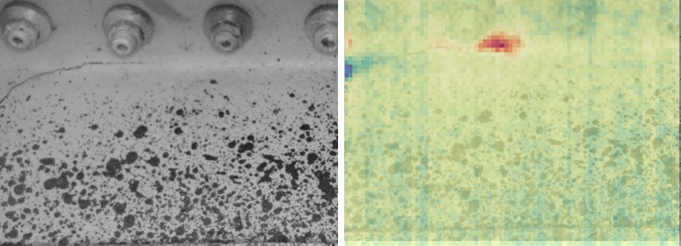 The image is a composite showing images from a visible camera (left) and processed data from infrared camera overlaid on the same visible image (right) from inside a wing box during a test at Empa with a crack extending from left to right with its tip surrounded by the red area in the right image. Each nut in the image is about 20 mm in diameter and a constant amplitude load at 1.25 Hz was being applied causing a wing tip displacement of 80 mm +/- 15 mm.
The University of Liverpool is the coordinator of the DIMES project and the other partners are Empa, Dantec Dynamics GmbH and Strain Solutions Ltd.
 The DIMES project has received funding from the Clean Sky 2 Joint Undertaking under the European Union’s Horizon 2020 research and innovation programme under grant agreement No. 820951.
The opinions expressed in this blog post reflect only the author’s view and the Clean Sky 2 Joint Undertaking is not responsible for any use that may be made of the information it contains.
References
[1] Burnham, R.S., McKinley, R.S. and Vincent, D.D., 2006. Three types of skin-surface thermometers: a comparison of reliability, validity, and responsiveness. American journal of physical medicine & rehabilitation, 85(7), pp.553-558.
[2] Middleton, C.A., Gaio, A., Greene, R.J. and Patterson, E.A., 2019. Towards automated tracking of initiation and propagation of cracks in aluminium alloy coupons using thermoelastic stress analysis. Journal of Nondestructive Evaluation, 38(1), p.18.

Pingback: My Engineering Day | Realize Engineering
Pingback: Noisy progressive failure of a composite panel | Realize Engineering