Over the last couple of years, we have been transitioning a technique, which we developed for tracking the motion of nanoparticles using caustics [see ‘Slow moving nanoparticles‘ on December 13th 2017], from its initial use in exploring mechanics at the nanoscale to applications in nanobiology [See ‘Label-free real-time tracking of individual bacterium‘ on January 25th, 2023] where it has the advantages of functioning in real-time and being label-free (chemical labels can impact motion, protein interactions and cell behaviour). In the summer, we had couple of articles published in consecutive issues of the Nature journal, Scientific Reports which describe our recent work. In the first, we have explored the diffusion of nanoparticles through a synthetic analogue of the vitreous humour in order to support the design of novel therapeutics for retinal diseases. Retinal diseases, such as macular degeneration and diabetic retinopathy, affects millions of people globally and treatment often involves frequent intravitreal injections of anti-vascular endothelium growth factor agents and corticoids. Delivery of the appropriate dose to the retinal cell layer is challenging due to the complex nature of the vitreous and functionalised nanoparticles offer a potential solution. In vivo animal testing is inappropriate because of the ethical concerns and poor representation of human eyes and ex vivo testing of cadaveric eyes is unreliable due to the instability of biomechanical and biochemical properties of the vitreous humour. Hence, we used agar-hyaluronic acid hydrogels as an in vitro model of the vitreous and employed the caustic technique to track the motion of nanoparticles through the hydrogels. The hydrogels had been validated as a representative model of the vitreous humour by other research groups. Our tracking technique revealed that the electric charge on the nanoparticles did not affect their diffusion through the hydrogel; however, both the diameter of the particles and the heterogeneous nature of the gel influenced the diffusion. Nanoparticles with diameters of 200, 100 and 50 nm moved progressively more quickly and over a larger area. The diffusion rates in hydrogels with a high viscosity (about 450 Pa.s) were consistent throughout the gel implying that the gel was homogeneous, while gels with medium (about 40 Pa.s) to low (about 3 Pa.s) viscosity generated diffusion rates that were distributed bi-modally suggesting a heterogeneous gel with zones of low and high density in which the particles moved more or less freely. The heterogeneity of a gel renders a global value for viscosity somewhat meaningless and makes comparisons difficult with the vitreous humour because it is also heterogeneous; however, global values of viscosity for porcine vitreous humour are typically 1 Pa.s. We are continuing this research; however, our published work has demonstrated that the use of caustics in an optical microscope is a reproducible and inexpensive technique for exploring the design of novel nanoscale drug delivery systems for the eye.
Source: Lorenzo Lopez M, Kearns VR, Curran JM, Patterson EA. Diffusion of nanoparticles in heterogeneous hydrogels as vitreous humour in vitro substitutes. Scientific reports. 2024 Jul 29;14(1):1744.
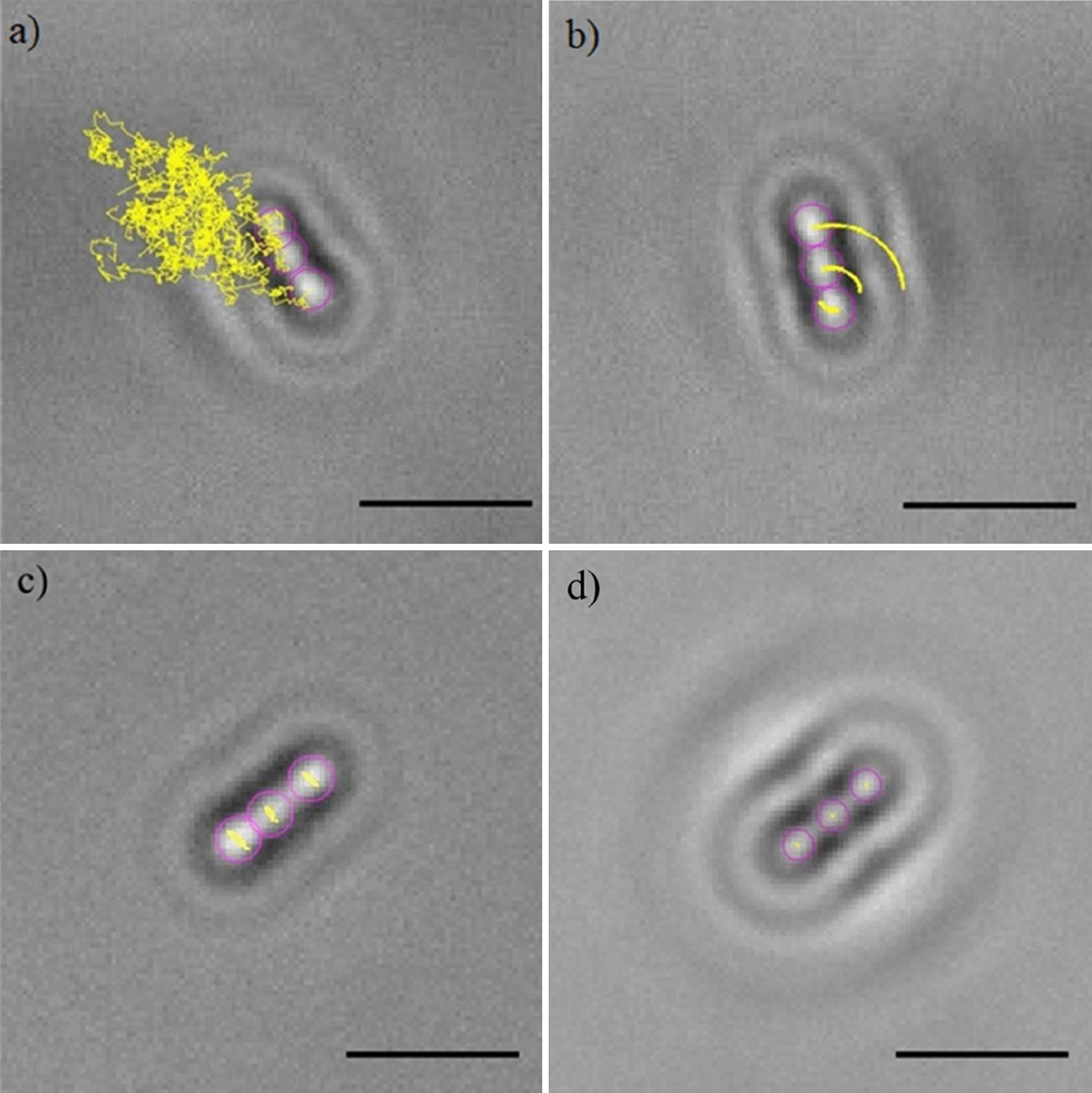
Image: Random track of a nanoparticle superimposed on its image generated in the microscope using a pin-hole and narrowband filter.