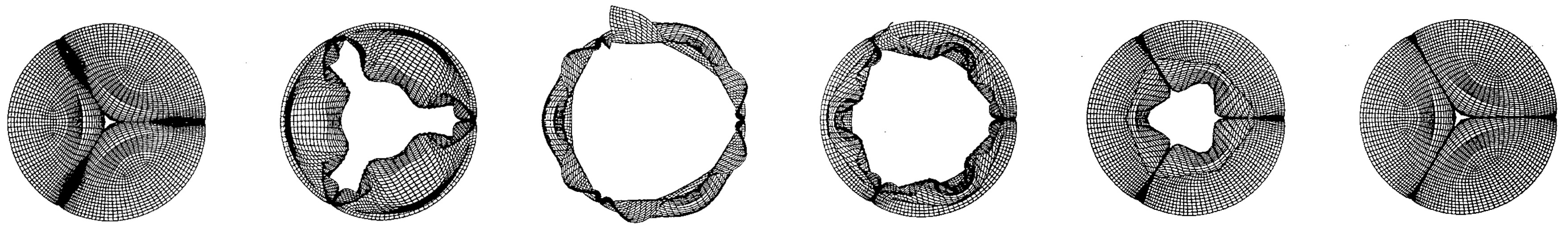
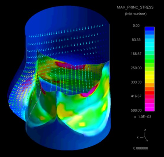
A couple of weeks ago I wrote about speaking to a workshop on the aorta and reminisced about research on cardiac dynamics from about 15 years ago. It triggered another memory of research we did more than 20 years ago on the tearing of the leaflets of artificial heart valves made from biological tissue. We developed a computational model of the stresses associated with a tear developing in a porcine bioprosthetic heart valve. The black and white images show snapshots of the predicted motion during the cardiac cycle of a damaged valve with a tear at about 11.30 along the edge of the top right leaflet. The valve was simulated as being implanted to replace the aortic valve and the view is from the aorta, i.e., looking in the opposite direction to the blood flow out of the heart. The tear causes part of the leaflet to flap outwards as can be seen in the middle snapshots. The colour image shows the distribution of stress in the leaflet corresponding to the last snapshot of the motion and the concentration of stress around the tip of the tear can be seen which will tend to cause the leaflet to tear further leading to a bigger flap, more regurgitation of blood. We were really excited about this research when we published it in 1999 but it has attracted relatively little attention in the last 23 years. I would like to think that we were far ahead of our times but that’s unlikely and probably it was not as exciting as we thought, maybe because it lacked clinical relevance, our model lacked credibility or not many people have found our paper.
Source: Chew GG, Howard IC & Patterson EA, Simulation of damage in a porcine prosthetic heart valve, J. Medical Engineering & Technology, 23(5):178-189, 1999.