What do marshmallows, jelly (or Jell-O), cream cheese and Chinese soup dumplings have in common? They are often made with gelatin. Gelatin is derived from the skin and bones of cattle and pigs through the partial hydrolysis of collagen. Gelatin is a physical hydrogel meaning that it consists of a three-dimensional network of polymer molecules in which a large amount of water is absorbed, as much as 90% in gelatin. These polymer molecules are cross-linked by hydrogen bonds, hydrophobic interactions and chain entanglements. External stimuli, such as temperature, can change the level of cross-linking causing the material to transition between its solid, liquid and gel states. This is why jelly sets in the fridge and melts when it’s heated up – the cross-links holding the molecules together break down. This type of responsive behaviour allows the properties of hydrogels to be controlled at the micro and sub-micron scale for a host of applications including tissue engineering, drug delivery, water treatment, wearable technologies, and supercapacitors. However, the design and manufacture of soft hydrogels can be challenging due to the lack of technology for measuring the local properties. Current quantitative techniques for measuring the properties of hydrogels usually focus on bulk properties and provide little data about local variations or real-time responses to external stimuli. My colleagues and I have used gold nanoparticles as probes in hydrogels to map the properties at the microscale of thermosensitive hydrogels undergoing real-time transition from the solid to gel phases [see ‘Passive nanorheological tool to characterise hydrogels’]. This is an extension, or perhaps more accurately an application, of our earlier work on tracking nanoparticles through the vitreous humour of the eye [see ‘Nanoparticle motion-through heterogeneous hydrogels’ on November 6th, 2024]. The novel technique, which yields passive nanorheological measurements, allows us to evaluate local viscosity, identify time-varying heterogeniety and monitor dynamic phase transitions at the micro through to nano scale. The significant challenges of other techniques, such as weak signals due to high water content and the dynamism of hydrogels, are overcome with a fast, inexpensive and user-friendly technology. Although, even with these advantages, you are unlikely to use it when you are making jelly or roasting marshmallows over the campfire; however, it is really useful for understanding the transport of drugs through biological hydrogels or designing manufacturing processes for artificial tissue.
Reference
Moira Lorenzo Lopez, Victoria R. Kearns, Eann A. Patterson & Judith M. Curran, Passive nanorheological tool to characterise hydrogels, Nanoscale, 2025,17, 15338-15347.
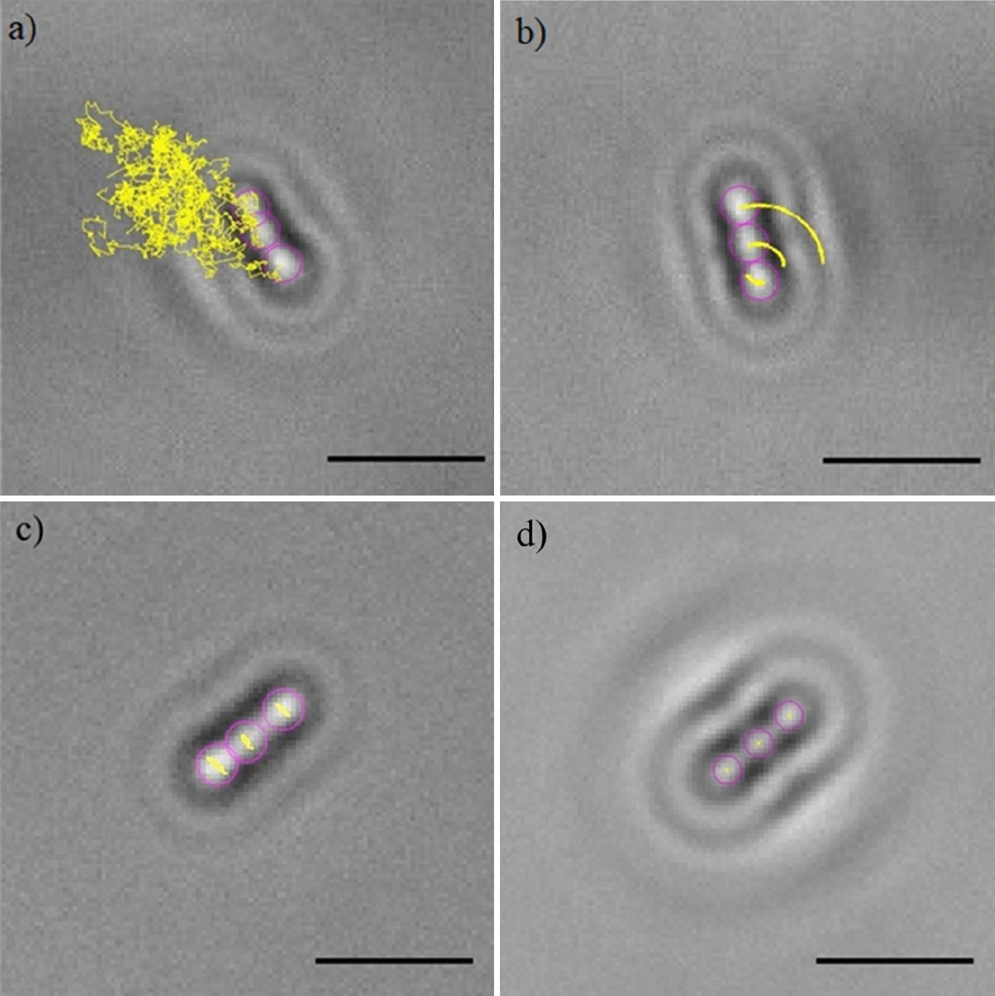
Image: Figure 5 from the above reference showing a hydrogel transitioning to a gel phase as result of an increase in temperature with 100 nm diameter gold nanoparticles with some particles (yellow arrows) at the interface between phases. The image was taken in an inverted optical microscope set up for tracking the nanoparticles.