Tag Archives: research

Reproducibility in science and technology

Schematic diagram from cited paper in Open Research Europe
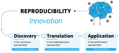
It has been suggested that there is crisis in science concerning the reproducibility of data [1].  New research findings are usually published based on data collected only by the group reporting the new findings, which raises the probability of bias in the results as well as reducing their likely validity.  It also creates a temptation to tamper with or falsify data given the incentives to publish.  It is unlikely that any prestigious journal would publish work that simply demonstrates that previously published findings can be reproduced consistently.  Yet, when they have tried to reproduce published data from experiments, many researchers have been unable to do so [2], which perhaps perversely makes the attempt to reproduce results publishable.  However, if no one has attempted to reproduce a published dataset then it stands until demonstrated to not be reproducible, which implies that much of the data in the published literature could be irreproducible and hence of dubious value.  This is a bigger problem than it might seem, because most scientific and technological innovation is built on the findings of fundamental research.  Hence, we are building on shaky foundations if results are not reproducible. Similarly, the transition from prototypes to reliable products is dependent on achieving reproducibility in the real-world of results obtained with a prototype in the laboratory.  I have been discussing these issues with a close collaborator for a number of years and last week we published a letter, in Open Research Europe, summarizing our views.  In ‘Achieving reproducibility in the innovation process’ [3], we propose that a different approach to reproducibility is required for each phase of the innovation process, i.e., discovery, translation and application, because reproducibility has different implications in each phase.  The diagram, reproduced from the paper (CC-BY-4.0), shows our ideas schematically but follow the link to read and comment on them.

References

[1] Baker, M. (2016). Reproducibility crisis. Nature, 533(26), 353-66.

[2] Camerer, C. F., Dreber, A., Holzmeister, F., Ho, T. H., Huber, J., Johannesson, M., … & Wu, H. (2018). Evaluating the replicability of social science experiments in Nature and Science between 2010 and 2015. Nature Human Behaviour, 2(9), 637-644.

[3] Whelan M & Patterson EA, (2025). Achieving reproducibility in the innovation process, Open Research Europe, 5:25. https://doi.org/10.12688/openreseurope.19408.1

Corona-induced transition from molecular to particle motion in biological media

Light signatures generated by particles in a nanoscopeIn last month’s post [see ‘Nanoparticle motion through heterogeneous hydrogels’ on November 6th, 2024], I described our recent work on tracking nanoparticles through a model of the vitreous humour and mentioned it was the first of two articles published in the Nature journal, Scientific Reports.  In the second article, we explored the use of caustics in an optical microscope [see ‘Seeing the invisible’ on October 29th, 2014] to track nanoparticles in biofluids.  Nanoparticles are below the resolution of an optical microscope because they are substantially smaller than the wavelength of visible light; hence, they are usually tracked using fluorescent markers or tags attached chemically to the nanoparticles.  These tags can influence both the motion of the particles and biological activity so caustics provide a label-free technique that allows particles to be tracked in real-time using a standard optical microscope.  In most of our previous research, we have tracked nanoparticles in transparent fluids such as water, glycerol-water mixtures, or the hydrogels described in last month’s post.  In our latest work, we have tracked small nanoparticles with diameters from 10 to 100 nm in common cell culture media with different concentrations of serum proteins.  These fluids are a ‘soup’ of complex protein molecules that interact with one another and the gold nanoparticles being tracked.  We found that the presence of proteins caused a reduction in the rate of diffusion for both positively- and negatively-charged particles and we concluded that the proteins form a corona around each nanoparticle effectively enlarging its diameter.  For larger nanoparticles, and those positively-charged, the enlargement appears to cause a transition from molecular motion, in which particle diameter is unimportant, to particle motion where larger particles diffuse more slowly.  We first explored this transition from fractional to classical Stokes-Einstein behaviour in simple fluids in 2017 [‘Slow moving nanoparticles‘ on December 13th 2017] and it seems likely to be complicated in these complex fluids.  Hence, understanding protein dynamics as well nanoparticle dynamics will be essential to the development of nanotechnologies applicable in biological environments.  So, we have lots more work to do!

Sources:

Schleyer G, Patterson EA, Curran JM. Label free tracking to quantify nanoparticle diffusion through biological media. Scientific Reports. 2024 Aug 13;14(1):18822.

Coglitore D, Edwardson SP, Macko P, Patterson EA, Whelan MP, Transition from fractional to classical Stokes-Einstein behaviour in simple fluids, Royal Society Open Science, 4:170507, 2017.

Nanoparticle motion through heterogeneous hydrogels

Over the last couple of years, we have been transitioning a technique, which we developed for tracking the motion of nanoparticles using caustics [see ‘Slow moving nanoparticles‘ on December 13th 2017], from its initial use in exploring mechanics at the nanoscale to applications in nanobiology [See ‘Label-free real-time tracking of individual bacterium‘ on January 25th, 2023] where it has the advantages of functioning in real-time and being label-free (chemical labels can impact motion, protein interactions and cell behaviour).  In the summer, we had couple of articles published in consecutive issues of the Nature journal, Scientific Reports which describe our recent work.  In the first, we have explored the diffusion of nanoparticles through a synthetic analogue of the vitreous humour in order to support the design of novel therapeutics for retinal diseases.  Retinal diseases, such as macular degeneration and diabetic retinopathy, affects millions of people globally and treatment often involves frequent intravitreal injections of anti-vascular endothelium growth factor agents and corticoids.  Delivery of the appropriate dose to the retinal cell layer is challenging due to the complex nature of the vitreous and functionalised nanoparticles offer a potential solution.  In vivo animal testing is inappropriate because of the ethical concerns and poor representation of human eyes and ex vivo testing of cadaveric eyes is unreliable due to the instability of biomechanical and biochemical properties of the vitreous humour.  Hence, we used agar-hyaluronic acid hydrogels as an in vitro model of the vitreous and employed the caustic technique to track the motion of nanoparticles through the hydrogels.  The hydrogels had been validated as a representative model of the vitreous humour by other research groups.  Our tracking technique revealed that the electric charge on the nanoparticles did not affect their diffusion through the hydrogel; however, both the diameter of the particles and the heterogeneous nature of the gel influenced the diffusion.  Nanoparticles with diameters of 200, 100 and 50 nm moved progressively more quickly and over a larger area.  The diffusion rates in hydrogels with a high viscosity (about 450  Pa.s) were consistent throughout the gel implying that the gel was homogeneous, while gels with medium (about 40 Pa.s) to low (about 3 Pa.s) viscosity generated diffusion rates that were distributed bi-modally suggesting a heterogeneous gel with zones of low and high density in which the particles moved more or less freely.  The heterogeneity of a gel renders a global value for viscosity somewhat meaningless and makes comparisons difficult with the vitreous humour because it is also heterogeneous; however, global values of viscosity for porcine vitreous humour are typically 1 Pa.s.  We are continuing this research; however, our published work has demonstrated that the use of caustics in an optical microscope is a reproducible and inexpensive technique for exploring the design of novel nanoscale drug delivery systems for the eye.

Source: Lorenzo Lopez M, Kearns VR, Curran JM, Patterson EA. Diffusion of nanoparticles in heterogeneous hydrogels as vitreous humour in vitro substitutes. Scientific reports. 2024 Jul 29;14(1):1744.

Image: Random track of a nanoparticle superimposed on its image generated in the microscope using a pin-hole and narrowband filter.

Extra on digital twins

After five months of posting monthly, I cannot resist the temptation to slip in an extra one.  Mainly because I want to let you know about the Pint of Science Festival taking place next week.  In Liverpool we have organised a series of three evenings at the Philharmonic pub on Hope Street featuring talks by engineers from the School of Engineering and the Institute for Digital Engineering and Autonomous Systems (IDEAS) at the University of Liverpool.  I am planning to talk about digital twins – what they are, how we can use them, what they might become and whether we are already part of a digital world.  If you enjoyed reading my posts on ‘Digital twins that thrive in the real world’, ‘Dressing up your digital twin’, and ‘Are we in a simulation?’ then come and discuss digital twins with me in person.  My talk is part of a programme on Digital with Everything on May 15th.  On May 13th and 14th we have programmes on Engineering in Nature and Science of Vision, Colliders and Crashes, respectively.  I hope you can come and join us in the real-world.