Tag Archives: forensic science

Press release!

A jumbo jet has about six million parts of which roughly half are fasteners – that’s a lot of holes.

It is very rare for one of my research papers to be included in a press release on its publication.  But that’s what has happened this month as a consequence of a paper being included in the latest series published by the Royal Society.  The contents of the paper are not earth shattering in terms of their consequences for humanity; however, we have resolved a long-standing controversy about why cracks grow from small holes in structures [see post entitled ‘Alan Arnold Griffith‘ on  April 26th, 2017] that are meant to be protected from such events by beneficial residual stresses around the hole.  This is important for aircraft structures since a civilian airliner can have millions of holes that contain rivets and bolts which hold the structure together.

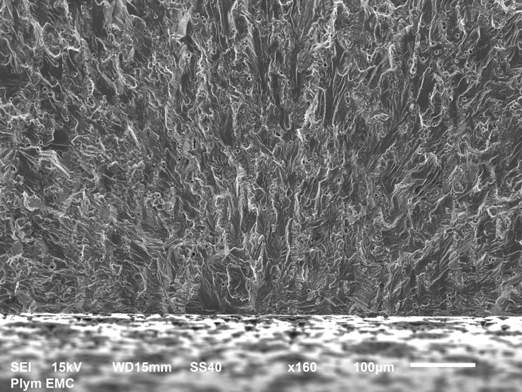
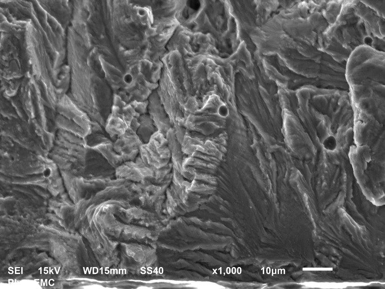
We have used mechanical tests to assess fatigue life, thermoelastic stress analysis to measure stress distributions [see post entitled ‘Counting photons to measure stress‘ on November 18th, 2015], synchrotron x-ray diffraction to evaluate residual stress inside the metal and microscopy to examine failure surfaces [see post entitled ‘Forensic engineering‘ on July 22nd, 2015].  The data from this diverse set of experiments is integrated in the paper to provide a mechanistic explanation of how cracks exploit imperfections in the beneficial residual stress field introduced by the manufacturing process and can be aided in their growth by occasional but modest overloads, which might occur during a difficult landing or take-off.

The success of this research is particularly satisfying because at its heart is a PhD student supported by a dual PhD programme between the University of Liverpool and National Tsing Hua University in Taiwan.  This programme, which supported by the two partner universities, is in its sixth year of operation with a steady state of about two dozen PhD students enrolled, who divide their time between Liverpool, England and Hsinchu, Taiwan.  The synchrotron diffraction measurements were performed, with a colleague from Sheffield Hallam University, at the European Synchrotron Research Facility (ESRF) in Grenoble, France; thus making this a truly international collaboration.

Source:

Amjad K, Asquith D, Patterson EA, Sebastian CM & Wang WC, The interaction of fatigue cracks with a residual stress field using thermoelastic stress analysis and synchrotron x-ray diffraction experiments, R. Soc. Open Sci. 4:171100.

Forensic engineering

Picture1The picture above shows the fracture surface of a thin bar of aluminium alloy that had a circular hole through the middle, like the peep-hole in a front door. The photograph was taken in a Scanning Electron Microscope (SEM) at x160 magnification. There is a scale bar in the bottom right corner showing a length of 100 microns. We are looking approximately in the longitudinal direction, which was the direction of loading, and across the photograph from left to right corresponds to the direction you would look through the hole. The lower one third of the picture shows the machined surface of the hole cut or machined by the drill. The top two-thirds shows the surface created by the fatigue crack as it extended incrementally with each cycle of load. The crack started from edge of the machined surface approximately on the vertical centre-line of the picture. I can tell this because all of the features in the texture of the fracture surface point towards this point because the failure radiated out from this location. The picture below shows the crack initiation area at x1000 magnification. It is a small area at interface with hole above the letters ‘SS40’ in the top photograph; this should be enough to let you identify the common features but the interpretation of these images requires significant skill.

Fractography is the forensic study of failure surfaces such as this to establish the cause of failure. In this example, the hole in aluminium bar ensured that it will always fail with cyclic loading through the growth of a crack from somewhere around the hole. The textured form of the fracture surface occurs because the material is not homogeneous at this scale but made up of small grains. The failure of each grain is influenced by its orientation to the loading which results in the multi-faceted surface in the photographs.

I made the photographs the focus of this post because I thought they are interesting, but may be that’s because I’m an engineer, and because they are a tiny part in a fundamental research programme on which I have been spending a significant portion of my time. A goal of programme is to understand how to use these materials to build more energy-efficient structures that are cheaper and last longer without failing by, for example, fatigue.

More details:

The bar was 1.6mm thick and 38mm wide in the transverse direction and made from 2024-T3 Aluminium alloy. The hole diameter was 6.36mm. A tension load was repeatedly applied and removed in the longitudinal direction which caused the initiation and growth of a fatigue crack from the hole that after many cycles of loading led to the bar breaking in half along a plane perpendicular to the load direction. The pictures were taken at the University of Plymouth by Khurram Amjad with the assistance of Peter Bond and Roy Moate using a JEOL JSM-6610LV.

x1000