Entropy management for bees and flights

entropy_vectorEngineers like to apply the second law of thermodynamics to chemical processes and power generation cycles. However, it has some useful lessons for everyday life since it can be paraphrased as ‘whenever you organise any process expect some disorder, or entropy to be generated’, so a shrewd person plans for disorder and designs in a bit of slack or redundancy.

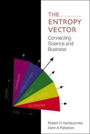
Bob and I gave an example of this in our book, ‘The Entropy Vector’.  We pointed out that if you plan your flight schedule to use all of the available gates at an airport then you will have unhappy passengers when flights are delayed, unless you plan for buses to unload planes parked away from the terminal. European airports tend to be good at this whereas US ones tend to leave passengers in planes that are unable to dock at the terminal.

Our example was inspired by frustrating experiences when we were writing the book. A more topical and important example was raised by Mark Winston in the New York Times on July 14th, 2014 in reporting the importance of bees to farming. His research team found that crop yields were maximised when large acreages were left uncultivated to support wild pollinators. He postulated that a variety of wild plants means a healthier, more diverse bee population which will be more active in the planted fields next door. Their numbers were startling with profits more than doubling for farmers that left a third of their acreage fallow. Winston highlights that this contravenes conventional wisdom that bees and fields can be micromanaged.

This seems like reinventing the wheel because I remember being taught about the importance of crop rotation, including a fallow period, in my ‘middle’ school geography classes. Oh dear, now I am showing my age.

The bottom-line is don’t micromanage. Allow for a bit of inefficiency, not too much of course or your competitors will get ahead! It’s a question of balance.

Seeing the invisible

Track of the Brownian motion of a 50 nanometre diameter particle

Track of the Brownian motion of a 50 nanometre diameter particle in a fluid.

Nanoparticles are being used in a myriad of applications including sunscreen creams, sports equipment and even to study the stickiness of snot!  By definition, nanoparticles should have one dimension less than 100 nanometres, which is one thousandth of the thickness of a human hair.  Some nanoparticles are toxic to humans and so scientists are studying the interaction of nanoparticles with human cells.  However, a spherical nanoparticle is smaller than the wavelength length of visible light and so is invisible in a conventional optical microscope used by biologists.  We can view nanoparticles using a scanning electron microscope but the electron beam damages living cells so this is not a good solution.  An alternative is to adjust an optical microscope so that the nanoparticles produce caustics [see post entitled ‘Caustics’ on October 15th, 2014] many times the size of the particle.  These ‘adjustments’ involve closing an aperture to produce a pin-hole source of illumination and introducing a filter that only allows through a narrow band of light wavelengths.  An optical microscope adjusted in this way is called a ‘nanoscope’ and with the addition of a small oscillator on the microscope objective lens can be used to track nanoparticles using the technique described in last week’s post entitled ‘Holes in liquid‘.

The smallest particles that we have managed to observe using this technique were gold particles of diameter 3 nanometres , or about 1o atoms in diameter dispersed in a liquid.


Image of 3nm diameter gold particle in a conventional optical microscope (top right), in a nanoscope (bottom right) and composite images in the z-direction of the caustic formed in the nanoscope (left).

Image of 3nm diameter gold particle in a conventional optical microscope (top right), in a nanoscope (bottom right) and composite images in the z-direction of the caustic formed in the nanoscope (left).

Sources:

http://ihcp.jrc.ec.europa.eu/our_activities/nanotechnology/jrc-scientists-develop-a-technique-for-automated-three-dimensional-nanoparticle-tracking-using-a-conventional-microscope

‘Scientists use gold nanoparticles to study the stickiness of snot’ by Rachel Feldman in the Washington Post on October 9th, 2014.

J.-M. Gineste, P. Macko, E.A. Patterson, & M.P. Whelan, Three-dimensional automated nanoparticle tracking using Mie scattering in an optical microscope, Journal of Microscopy, Vol. 243, Pt 2 2011, pp. 172–178

Patterson, E.A., & Whelan, M.P., Optical signatures of small nanoparticles in a conventional microscope, Small, 4(10): 1703-1706, 2008.

Holes in fluids

Out-of-focus image from optical microscope of 10 micron diameter polystrene spheres in water

Out-of-focus image from optical microscope of 10 micron diameter polystyrene spheres in water

The holes that I wrote about last week and the week before (post entitled ‘Holes‘ on October 8th)were essentially air-filled holes in a solid plate.  When an in-plane load is applied to the plate it deforms and its surface around the hole becomes curved due to the concentration of stress and light passing through the curved surfaces is deviated to form the caustic.  If you didn’t follow that quick recap on last week then you might want flip back to last week’s post before pressing on!

The reverse situation is a solid in a fluid.  It is difficult to induce stress in a fluid so instead we can use a three-dimensional hole, i.e. a sphere, to generate the curve surface for light to pass through and be deviated.  This is quite an easy experiment to do in an optical microscope with some polystyrene spheres floating in distilled water with the microscope slightly out of focus you get bright caustics.  And if you take a series of photographs (the x-y plane) with the microscope objective lens at different heights (z-value) it is possible to reconstruct the three-dimensional shape of the caustic by taking the intensity or greyscale values along the centre line of each image and using them all to create new image of the x-z and, or y-z plane, as shown in the picture.

Well done if you have got this far and are still with me!  I hope you can at least enjoy the pictures.  By the way the particle in the images is about the same diameter as a human hair.

Image in optical microscope of polystrene particle in water (left), series of images at different positions of microscope objective (centre) and artificial image created from greyscale data along centre-lines of image series (right).

Image in optical microscope of polystyrene particle in water (left), series of images at different positions of microscope objective (centre) and artificial image created from greyscale data along centre-lines of image series (right).

Source:

Patterson, E.A., & Whelan, M.P., Tracking nanoparticles in an optical microscope using caustics, Nanotechnology, 19(10): 105502, 2008.

Caustics

caustic_hole

White light caustic of 4mm diameter hole in 6mm (PMMA) plate subject to 3kN tension

As children many of us have burnt a hole (yes, tenuous link to last week’s post on ‘Holes’) in a piece of paper by focussing the sun’s rays with a magnifying glass. If you move the glass up or down and tilt it slightly then the sun’s rays will not be focussed on a spot and instead you see a complex spiralling pattern of light. This pattern is caused by the rays being bent by their passage through different sections of the curved glass. The same type of pattern, known as a caustic, appears on the bottom of your bath when you let (clean) water run out down the plug-hole if you have spotlights above the bath. This caustic is produced by the light rays from the spotlight being bent by varying degrees depending on where they pass through the vortex formed by the water spinning down the hole.  Caustics can also be produced when light passes through a glass of water or on the bottom of an outdoor swimming pool in bright sunlight.

The top picture shows the caustic formed by light passing through a transparent plate with a hole when the plate is stretched in the vertical direction. The load in the plate has to flow around the hole where it ‘bunches up’ or concentrates (see last week’s post entitled ‘Holes’) which causes high levels of local deformation with the plate thinning non-linearly at the intersection of the hole circumference and horizontal diameter. When the light passes through the deformed region it is deviated by amount dependent on the local thinning and forms the pattern shown.

This is not a totally abstract phenomenon because the same mechanism of thinning occurs at the tip of cracks as a result of the very high stress concentration at the sharp crack tip, as shown schematically in the diagram below. So we can evaluate the stress concentration by measuring the caustic it generates; it is even possible to predict in which direction the crack will grow next.

Schematic diagram of transparent plate with a crack loaded vertically in tension (left), light ray tracings through the cracked region (centre) and caustic formed on a screen (right).

Schematic diagram of transparent plate with a crack loaded vertically in tension (left), light ray tracings through the cracked region (centre) and caustic formed on a screen (right).

For information:

Carazo-Alvarez, J.D., Patterson, E.A., 1999, ‘A general method for automated analysis of caustics’, Optics & Lasers in Engng., 32: 95-110.

http://lgg.epfl.ch/caustics/